Leg Anatomy Muscles Ligaments And Tendons : Benefit Pt S Anatomy Series The Ankle. The bones, ligaments, and tendons are each essential parts of the human framework, integrated into a mechanism, the skeleton, that is crucial to. Collectively, they act to dorsiflex and invert the foot at the ankle joint. The tendons of the edl can be palpated on the dorsal surface of the foot. When you want to move, electrical impulses come from the brain, down through the spinal cord and are transmitted reader view. Maximize performance & minimize injuries. he can be found on.
When everything works together, the ankle functions. They are the continuations of muscles and. The muscles of the thigh and lower leg are comprised of compartments defined as distinct anatomical spaces bordered by fascia or bone. As with any structure, the human body is built upon a framework that is constructed to carry out a wide range of functions. Muscles, either individually or in groups, are supported by fascia.
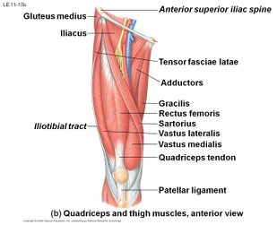
Quadriceps Tendon Tear Physiopedia from www.physio-pedia.com Tendons consist of densely packed collagen fibers. As with any structure, the human body is built upon a framework that is constructed to carry out a wide range of functions. Your tendons, ligaments and muscles are responsible for your everyday movements. The muscles, tendons, and ligaments that support the ankle joint work together to propel the body. Want to learn more about it? Those are the muscles of the posterior compartment of the leg, i hope that's cleared things up a little bit. Ligaments also support the lower end of the leg where it forms a hinge for the ankle. The patellar tendon on the front of the knee is part of the quadriceps mechanism.
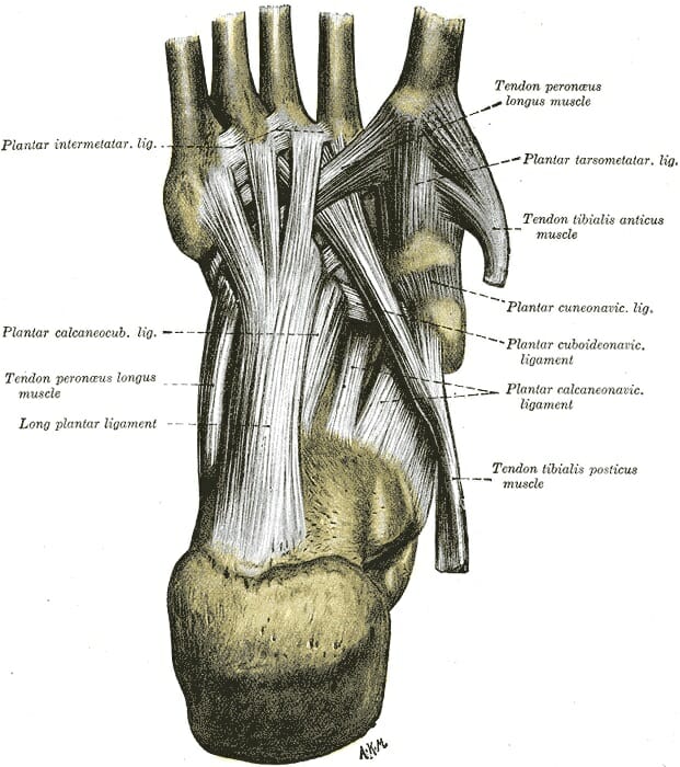
Muscles, tendons, and ligaments run along the surfaces of the feet, allowing the complex movements needed for motion and balance.
Tendons connect muscles to bones. Click now to learn more about the bones, muscles, and soft tissues of leg and knee anatomy: Collectively, they act to dorsiflex and invert the foot at the ankle joint. Learn how they work together to avoid injury and stay active. Ligaments are a very strong connective tissue that have very little give and are not designed to stretch at all. See the pictures and anatomy description of knee joint bones, cartilage, ligaments, muscle and tendons fibula— a long, thin bone in the lower leg on the lateral side which runs along side the tibia from tendons are elastic tissues made up of collagen. We speak of the upper extremities (arms) and the lower extremities (legs). The human leg, in the general word sense, is the entire lower limb of the human body, including the foot, thigh and even the hip or gluteal region. Related online courses on physioplus. The patellar tendon on the front of the knee is part of the quadriceps mechanism. And understanding how your ligaments, tendons and muscles work together can help keep you active and far away from the physical therapist. Muscles, tendons, and ligaments run along the surfaces of the feet, allowing the complex movements needed for motion and balance. Tendons are not elastic by nature of their collagen fibril organizat.
When a muscle contracts, it exerts mechanical force on the tendon. Muscles, ligaments, & tendons by: Muscles, either individually or in groups, are supported by fascia. Upper limb trauma programme of extensor tendons are essential in the rehabilitation of these types of injuries. In addition, there are some other minor anatomical differences.
Foot Anatomy Bones Ligaments Muscles Tendons Arches And Skin from biologydictionary.net One way our muscles work: About halfway down the lower leg the muscle fibers merge into a broad flat tendon, which then the foot is a fascinating structure, composed of many bones, ligaments, and cartilages. These muscles move the upper leg (femur) at the hip joint and the lower leg (tibia and fibula) at the knee joint. Learn about the muscles, tendons, bones, and ligaments that comprise the knee joint anatomy. Maximize performance & minimize injuries. he can be found on. As you can see, the anatomy of the ankle is very complex. We speak of the upper extremities (arms) and the lower extremities (legs). As with any structure, the human body is built upon a framework that is constructed to carry out a wide range of functions.
Other smaller muscles and tendons surround the knee joint as well.
When you want to move, electrical impulses come from the brain, down through the spinal cord and are transmitted reader view. Skeletal muscles are held to the bones with the help of tendons. Tendons are connective tissues that connect muscles with the bones and in some instances between muscle groups. Смотреть все результаты для этого вопроса. Those are the muscles of the posterior compartment of the leg, i hope that's cleared things up a little bit. Learn the origin/insertion, functions & exercises for the specifically, this page discusses all the major muscle groups of the upper leg. The muscles, tendons, and ligaments that support the ankle joint work together to propel the body. Watch cervical muscle anatomy animation. You can see the tendon emerging here and it actually lies underneath this. Originates from the lateral condyle of the tibia and the medial surface of the fibula. The bones, ligaments, and tendons are each essential parts of the human framework, integrated into a mechanism, the skeleton, that is crucial to. In addition, there are some other minor anatomical differences. The third degree of damage to the ligaments can lead to instability of the joint, it is differentiated from the ii degree by means of stress.
Tendons connect muscles to bones, while ligaments connect bones to other bones. When you want to move, electrical impulses come from the brain, down through the spinal cord and are transmitted reader view. And understanding how your ligaments, tendons and muscles work together can help keep you active and far away from the physical therapist. Learn how they work together to avoid injury and stay active. As you can see, the anatomy of the ankle is very complex.
Leg Concise Medical Knowledge from cdn.lecturio.com The bones, ligaments, and tendons are each essential parts of the human framework, integrated into a mechanism, the skeleton, that is crucial to. Other smaller muscles and tendons surround the knee joint as well. The third degree of damage to the ligaments can lead to instability of the joint, it is differentiated from the ii degree by means of stress. Collectively, they act to dorsiflex and invert the foot at the ankle joint. Unlike tendons, which connect muscle to bone, ligaments connect bones to other bones. Unfortunately many of us live in a bodily environment where ligaments. Tendons of the lower leg, muscles tendons and ligaments of the upper leg. Maximize performance & minimize injuries. he can be found on.
Unfortunately many of us live in a bodily environment where ligaments.
Learn about the muscles, tendons, bones, and ligaments that comprise the knee joint anatomy. Ligaments are located at joints, whereas tendons provide the connection between muscle and bone that allows the muscles to move different parts of. Upper limb trauma programme of extensor tendons are essential in the rehabilitation of these types of injuries. These all work together to bear weight. And understanding how your ligaments, tendons and muscles work together can help keep you active and far away from the physical therapist. Maximize performance & minimize injuries. he can be found on. Originates from the lateral condyle of the tibia and the medial surface of the fibula. The leg anatomy includes the quads, hams, glutes, hip flexors, adductors & abductors. Related online courses on physioplus. Possible ruptures of ligaments, muscles and tendons. Muscles, ligaments, & tendons by: Tendons consist of densely packed collagen fibers. We speak of the upper extremities (arms) and the lower extremities (legs).
Leg Anatomy Muscles Ligaments And Tendons : Benefit Pt S Anatomy Series The Ankle. There are any Leg Anatomy Muscles Ligaments And Tendons : Benefit Pt S Anatomy Series The Ankle in here.